
Light Sheet Microscopy
Light sheet fluorescence microscopy (LSFM) has emerged as a transformative technique in the field of biological imaging, offering unprecedented capabilities for visualizing and quantifying dynamic processes within living organisms and tissues. This non-invasive, high-speed imaging method utilizes a thin sheet of light to selectively illuminate a sample, enabling optical sectioning and minimizing phototoxicity and photobleaching compared to traditional fluorescence microscopy techniques.
To comprehend LSM’s operation, two key components must be considered. The first is the creation and orientation of the light sheet, which is crucial for selective illumination. The second is the detection system that captures the fluorescence emitted from the sample.
Creating a Light Sheet
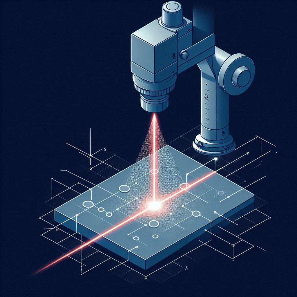
In light sheet fluorescence microscopy (LSFM), a thin sheet of light is used to selectively illuminate a sample, enabling optical sectioning and minimizing phototoxicity compared to traditional fluorescence microscopy techniques. The light sheet is typically generated by focusing a laser beam into a cylindrical lens, which compresses the beam in one dimension while leaving it unchanged in the orthogonal direction. This results in an elliptical beam profile with a narrow waist, known as the light sheet. The sample is positioned at the waist of the light sheet, where the illumination is most intense and the sheet is thinnest, ensuring that only a thin slice of the sample is excited. The fluorescence emitted from the illuminated plane is then collected by a wide-field detection objective, which is oriented perpendicular to the light sheet. This orthogonal arrangement of the illumination and detection pathways is a key feature of LSFM, as it allows for intrinsic optical sectioning and minimizes out-of-focus light.
In contrast, traditional fluorescence microscopy techniques, such as widefield and confocal microscopy, rely on illuminating the entire sample or a focused spot, respectively, leading to significant out-of-focus light and increased photobleaching and phototoxicity. By selectively illuminating only the plane of interest, LSFM reduces the overall exposure of the sample to light, enabling long-term, high-speed, and minimally invasive imaging of living specimens. The thin light sheet also ensures that the excitation light is confined to the focal plane of the detection optics, further improving the signal-to-noise ratio and contrast of the acquired images.

Figure 1. Simplified diagram showing a light sheet passing through some sample and exciting fluorescence.
Detection System
The detection system is designed to efficiently collect the fluorescence signal from the illuminated plane while minimizing out-of-focus light. This is achieved by using a wide-field detection objective oriented perpendicular to the light sheet, creating an orthogonal arrangement between the illumination and detection pathways. The fluorescence emitted from the illuminated plane is captured by the detection objective and projected onto a camera sensor.
Laser Systems in LSFM
The laser system used in LSFM plays a crucial role in generating the light sheet. Coherent lasers, such as diode-pumped solid-state lasers or gas lasers, are commonly employed due to their high spatial coherence, which allows for efficient focusing and shaping of the beam. The high temporal coherence of these lasers also enables the use of interference patterns to create structured illumination, which can be used for enhanced contrast or resolution.
The power of the laser is an important consideration, as it determines the intensity of the light sheet and the resulting fluorescence signal. Higher laser power can lead to increased signal-to-noise ratio but may also cause excessive photobleaching or phototoxicity. The choice of laser wavelength depends on the fluorophores used in the sample and the desired penetration depth, with longer wavelengths generally providing better tissue penetration.
Advantages of LSM
The key difference between the LSFM detection system and that of confocal or widefield microscopy lies in the selective illumination of the sample. In confocal microscopy, a focused laser beam scans the sample point-by-point, and a pinhole is used to reject out-of-focus light, improving the axial resolution. However, this point-scanning approach is inherently slower than the wide-field detection used in LSFM. Widefield microscopy, on the other hand, illuminates the entire sample, leading to significant out-of-focus light and reduced contrast. In contrast, LSFM illuminates only a thin slice of the sample with the light sheet, ensuring that the fluorescence signal collected by the detection objective originates predominantly from the focal plane. This selective illumination, combined with the wide-field detection, allows for rapid acquisition of entire planes, enabling high-speed volumetric imaging with minimal out-of-focus light and reduced photobleaching. The orthogonal arrangement of the illumination and detection pathways also provides inherent optical sectioning, eliminating the need for physical sectioning of the sample. The efficient detection system in LSFM, coupled with the selective illumination, enables the acquisition of high-contrast, low-background images at high speeds, making it a powerful tool for studying dynamic biological processes in living specimens.
In summary, Light Sheet Microscopy stands out as a powerful tool in biological research, providing scientists with detailed 3D images while preserving the integrity of their delicate samples. At Integrated Optics we manufacture a multitude of lasers suitable for Light Sheet Microscopy. They maintain a small form factor without compromising on performance, enabling manufacturers and researchers to integrate them in their LSFM systems.
Relevant Products
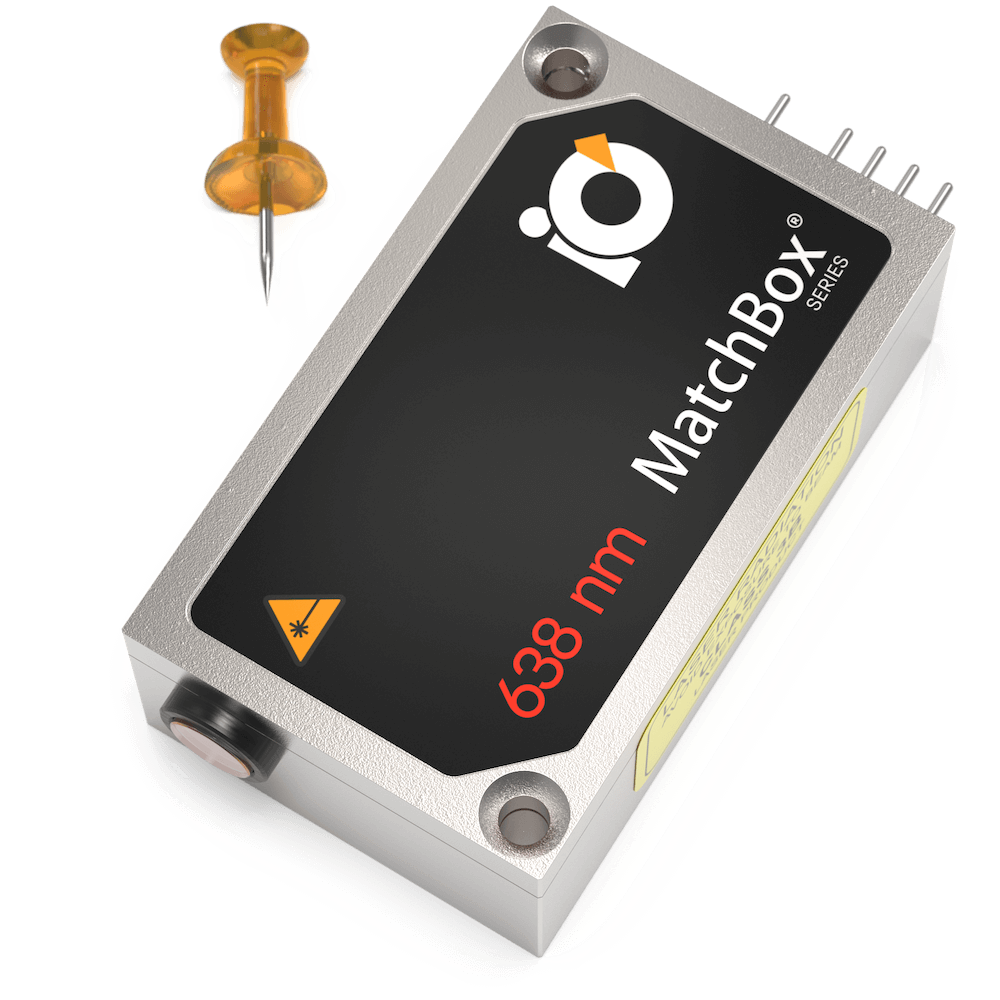
638 nm SLM Laser
Spectral line width FWHM, MHz: 2
Output power, mW: 60
Power stability, % (RMS, 8 hrs): 0.1
Intensity noise, % (RMS, 20 Hz to 20 MHz): 0.25
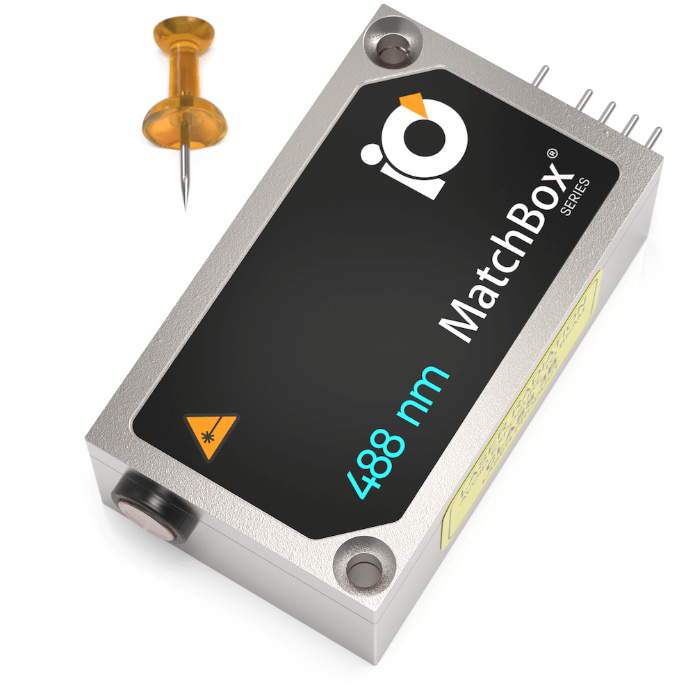
488 nm SLM Laser
Spectral line width FWHM, pm: 0.1
Output power, mW: 10
Power stability, % (RMS, 8 hrs): 0.2
Intensity noise, % (RMS, 20 Hz to 20 MHz): 0.2

Multi-Wavelength Laser
450 nm - 50
520 nm - 70
638 nm - 100
Power stability, % (RMS, 8 hrs): 0.2
Spectral line width FWHM, nm: <1.5
Intensity noise, % (RMS, 20 Hz to 20 MHz): <1

Multi-Wavelength Laser
488 nm - 40
520 nm - 80
638 nm - 130
Spectral line width FWHM, nm: 1
Power stability, % (RMS, 8 hrs): 0.2
Intensity noise, % (RMS, 20 Hz to 20 MHz): 0.5
Multi-Wavelength Laser
488 nm - 40
520 nm - 70
638 nm - 100
Spectral line width FWHM, nm: 1
Power stability, % (RMS, 8 hrs): 0.2
Intensity noise, % (RMS, 20 Hz to 20 MHz): 0.5
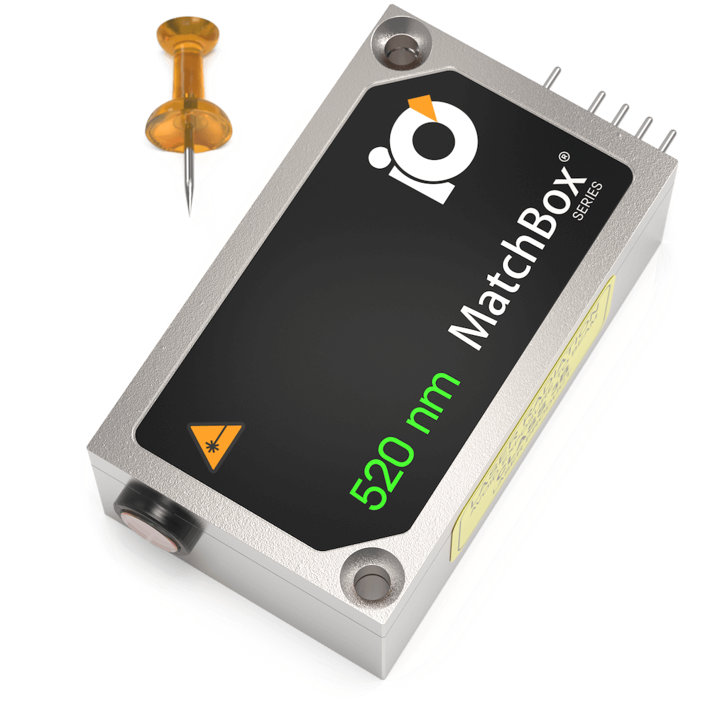
520 nm SLM Laser
Spectral line width FWHM, pm: 0.1
Output power, mW: 15
Power stability, % (RMS, 8 hrs): 0.05
Intensity noise, % (RMS, 20 Hz to 20 MHz): 0.5
488 nm SLM Laser
Spectral line width FWHM, pm: 0.1
Output power, mW: 30
Power stability, % (RMS, 8 hrs): 0.05
Intensity noise, % (RMS, 20 Hz to 20 MHz): 0.1
520 nm SLM Laser
Spectral line width FWHM, pm: 0.1
Output power, mW: 40
Power stability, % (RMS, 8 hrs): 0.05
Intensity noise, % (RMS, 20 Hz to 20 MHz): 0.5
406 nm SLM Laser
Spectral line width FWHM, MHz: 20
Output power, mW: 100
Power stability, % (RMS, 8 hrs): 0.05
Intensity noise, % (RMS, 20 Hz to 20 MHz): 0.2
638 nm SLM Laser
Spectral line width FWHM, MHz: 2
Output power, mW: 100
Power stability, % (RMS, 8 hrs): 0.03
Intensity noise, % (RMS, 20 Hz to 20 MHz): 0.25
Multi-Wavelength Laser
Power stability, % (RMS, 8 hrs): 0.2
Intensity noise, % (RMS, 20 Hz to 20 MHz): 0.5

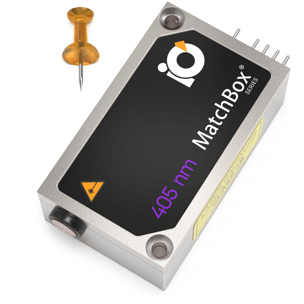
405 nm SLM Laser
Spectral line width FWHM, MHz: 20
Output power, mW: 50
Power stability, % (RMS, 8 hrs): 0.1
Intensity noise, % (RMS, 20 Hz to 20 MHz): 0.2

405 nm SLM Laser
Spectral line width FWHM, MHz: 20
Output power, mW: 100
Power stability, % (RMS, 8 hrs): 0.05
Intensity noise, % (RMS, 20 Hz to 20 MHz): 0.2