Applications

Atmospheric LiDAR
Atmospheric LiDAR is an advanced remote sensing technology that employs laser beams to analyze the Earth's atmosphere. By measuring the backscattered light from atmospheric particles, it provides detailed information on vertical profiles, aiding meteorology, climate research, and air quality monitoring. Atmospheric LiDAR plays a pivotal role in understanding complex atmospheric processes and contributes essential data for climate models and environmental assessments.

Flash LiDAR
Flash LiDAR is an advanced remote sensing technology that utilizes laser pulses in a rapid, sweeping fashion to create high-resolution 3D maps of the surroundings. Unlike traditional Lidar systems that rely on scanning mechanisms, Flash Lidar captures the entire scene simultaneously, providing real-time and comprehensive spatial information. This innovative approach enhances the speed and efficiency of mapping, making Flash Lidar well-suited for applications in autonomous vehicles, robotics, and environmental monitoring.
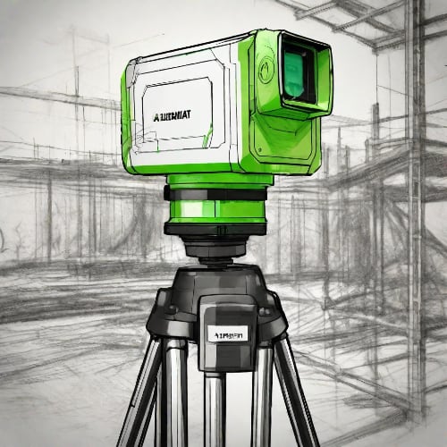
Light Detection and Ranging (LiDAR)
LiDAR (stands for Light Detection and Ranging) is a remote measurement technique, that incorporates laser pulses (used in most types of LiDAR) or laser frequency modulation for distance estimation and scanning of environment. The technology of LiDAR is built on basic principles of physics – the range measurement is calculated by taking into account the time it took for the laser pulse to reflect off of a surface and echo back to a detector or a sensor.

Stationary Terrestrial LiDAR
Stationary Terrestrial LiDAR, also known as Static Terrestrial LiDAR, is a technology used for non-contact 3D mapping and monitoring of stationary objects and environments. Unlike traditional mobile LiDAR systems mounted on vehicles, stationary LiDAR setups are fixed in a specific location. These systems utilize laser beams emitted from a stationary unit to measure distances and create detailed 3D point clouds of the surrounding area.

Confocal Microscopy
Confocal microscopy is a powerful imaging technique used in biological and materials science research. By employing point illumination and a spatial pinhole, confocal microscopy eliminates out-of-focus light, resulting in sharper, high-resolution images. This method enables three-dimensional imaging of specimens with exceptional optical sectioning, making it valuable for studying biological structures and dynamic processes at the cellular and subcellular levels.
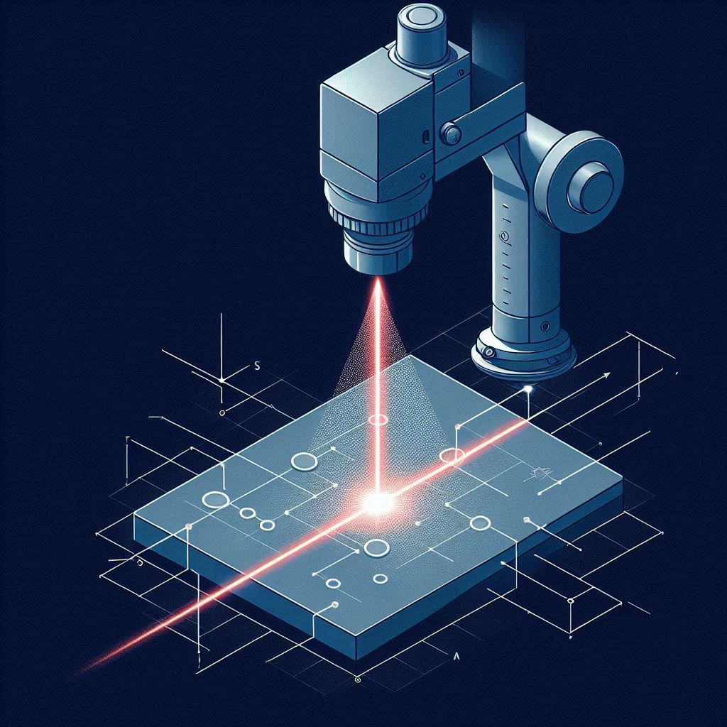
Light Sheet Microscopy
Light sheet fluorescence microscopy (LSFM) has emerged as a transformative technique in the field of biological imaging, offering unprecedented capabilities for visualizing and quantifying dynamic processes within living organisms and tissues. This non-invasive, high-speed imaging method utilizes a thin sheet of light to selectively illuminate a sample, enabling optical sectioning and minimizing phototoxicity and photobleaching compared to traditional fluorescence microscopy techniques.
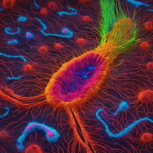
Super-resolution Microscopy
Traditional microscopy methods have served researchers for decades. Most conventional microscopes can image at resolutions between 1µm and 250nm which is sufficient to observe cellular dynamics and many internal structures. However, the necessity to discern finer details and develop new molecular based therapeutics has lead to the field of Super-Resolution Microscopy.

Total Internal Reflection Fluorescence (TIRF) Microscopy
TIRF microscopy is a powerful optical technique used primarily in the field of cell biology. It allows for the observation of phenomena occurring at the interface between a cell and a substrate, such as cell adhesion, membrane dynamics, and signal transduction events.
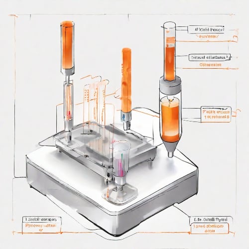
Flow Cytometry
Flow cytometry is a sophisticated analytical technique widely used in biomedical research and clinical diagnostics. It allows for the simultaneous analysis of multiple physical and chemical characteristics of cells or particles as they flow through a laser beam. By utilizing fluorescence and light-scattering principles, flow cytometry provides valuable insights into cell populations, allowing researchers to study cell morphology, identify cell types, and assess various cellular functions with high-throughput precision.
Nitrogen-Vacancy Magnetometry
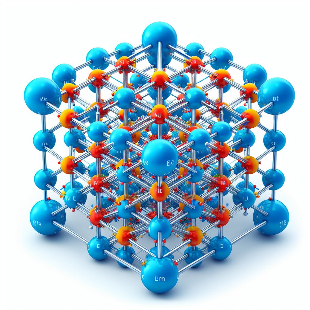
Nitrogen-Vacancy (NV) Magnetometry is a novel technique used to precisely measure the magnetic fields of a sample. It relies on an "imperfection" of a diamond's crystal lattice, where one pair of carbon-carbon bonds is replaced with a single nitrogen molecule. By forming a nitrogen-vacant cavity, also called a color center, unique properties of the valence electron can be utilised. Under the right conditions and with the help of lasers, the electron can become especially sensitive to magnetic fields. This property yields extraordinary responsiveness even under ambient conditions and allows researchers to explore nanoscale structures and processes in their sample.

Quantum Cryptography
Quantum cryptography is a way of securing information using the principles of quantum physics. One method of quantum cryptography is quantum key distribution (QKD), which allows two parties to share a secret key that can encrypt and decrypt messages. QKD uses entangled photons, which are pairs of light particles that have a quantum connection and share the same properties. By measuring the polarization of one photon, the other photon will have the same polarization, even if they are far apart. This way, the two parties can generate a random sequence of bits that form the key. However, to create and send entangled photons from space, they need small lasers that can fit into smallsats.
Fluorescence Lifetime Decay
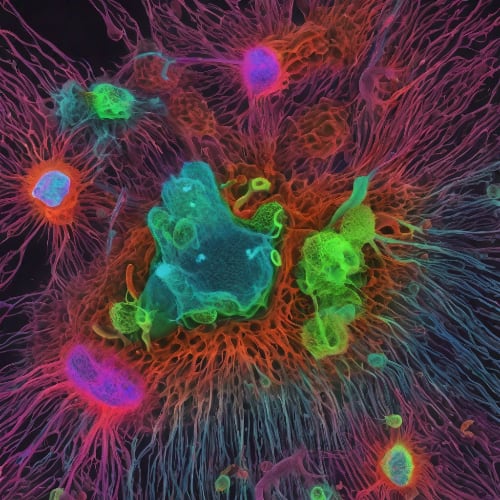
Fluorescence lifetime decay is the process of measuring how long a fluorescent molecule stays in its excited state before returning to its ground state. It can provide information about the molecular environment and interactions of the fluorescent molecule, as well as its structure and dynamics.
Fluorescence Spectroscopy
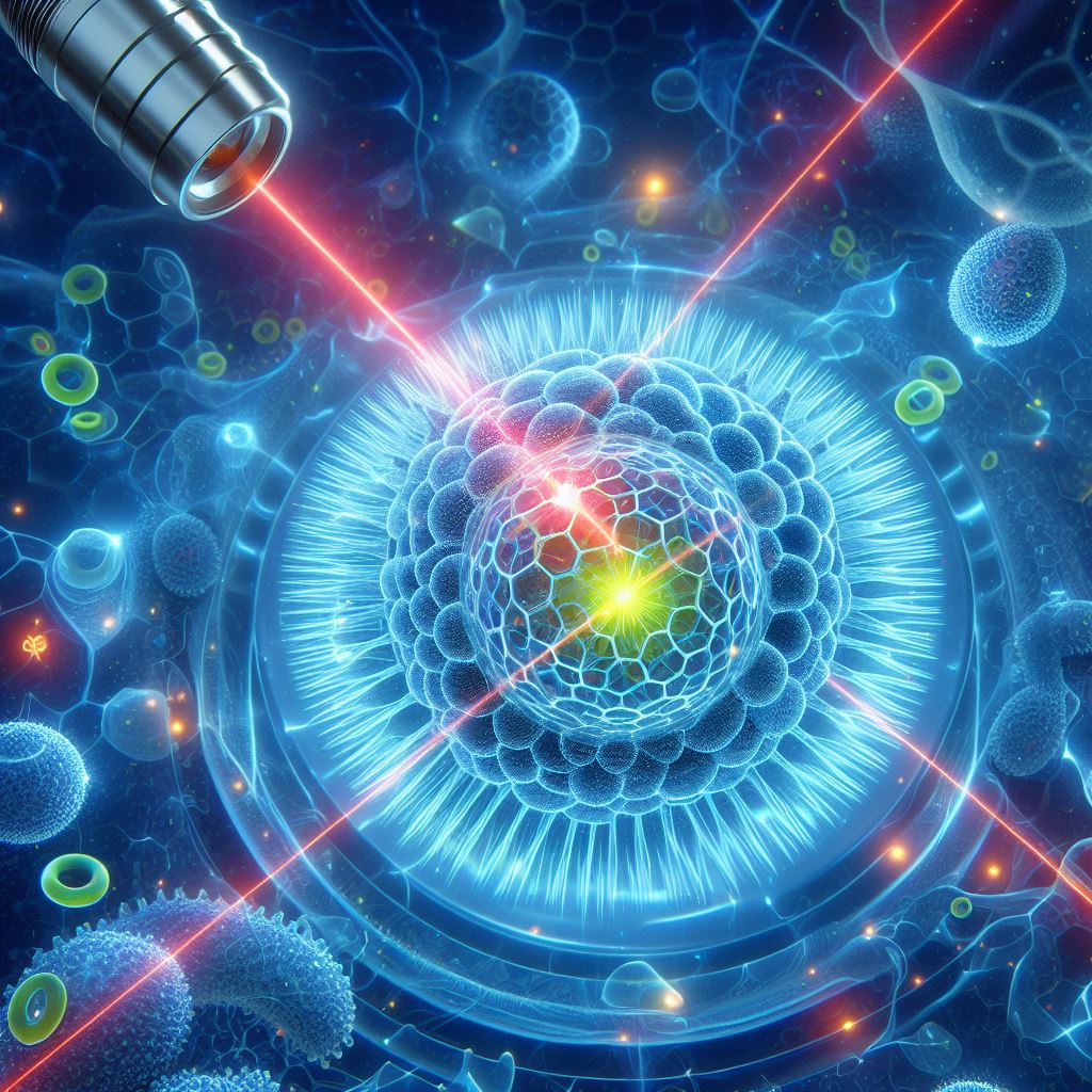
Fluorescence spectroscopy, with its origins in the early 20th century, has evolved into a sophisticated tool widely used in analytical chemistry, molecular biology, and pharmacology. The prevalence of this tool and its diverse applications can be equally attributed to developments in optics and laser technology as much as breakthroughs in the natural sciences.
Laser Induced Breakdown Spectroscopy (LIBS)
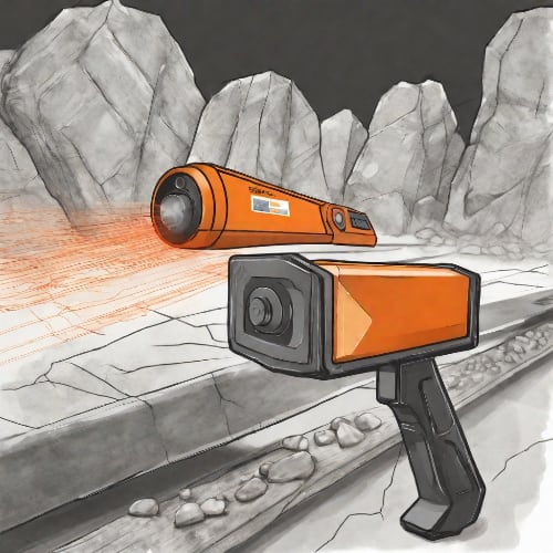
LIBS stands for laser-induced breakdown spectroscopy and is a rapid chemical analysis technique, first introduced in 1960s. Unlike in Raman spectroscopy, it uses short laser pulses to create plasma plumes on the surface of a sample and the spectrum is characterized of this plasma using low-cost spectrometers. It is also categorized as an in situ operation, that’s duration can last up to only a few seconds.
Raman Spectroscopy
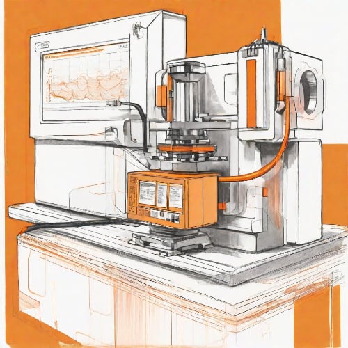
Raman Spectroscopy is a powerful analytical technique that explores molecular vibrations by measuring inelastic scattering of monochromatic light. It provides valuable insights into molecular structure, composition, and chemical bonding, making it widely used in material science, chemistry, and biology. The unique spectral fingerprints obtained through Raman spectroscopy enable non-destructive and precise identification of substances, making it a versatile tool for research and quality control applications.
Photoacoustic imaging
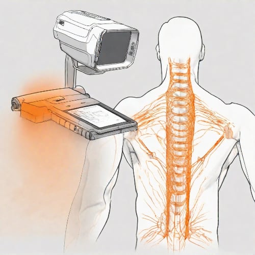
Photoacoustic imaging is a process where powerful laser pulses interact with material by exciting acoustic waves. Similarly, as in ultrasound imaging, the propagating acoustic waves are analyzed by piezo-based detectors (pick-ups), and the complete 3D image is formed by raster scanning or other techniques, such as laser-based holography or interferometry.