
Confocal Microscopy

Confocal microscopy is a powerful imaging technique used in biological and materials science research. By employing point illumination and a spatial pinhole, confocal microscopy eliminates out-of-focus light, resulting in sharper, high-resolution images. This method enables three-dimensional imaging of specimens with exceptional optical sectioning, making it valuable for studying biological structures and dynamic processes at the cellular and subcellular levels.
Confocal microscopes were built on the foundation laid by fluorescence microscopy. They are more complex instruments utilising the same principles, but capable of much more. Therefore, a brief overview of fluorescence microscopy will assist in understanding this topic.
Principle of a Fluorescence Microscope
Fluorescence microscopy is one of many fields encompassed by fluorescence spectroscopy. It utilizes the principle of fluorescence, which we have discussed extensively and have linked in this article. A basic fluorescence microscope usually uses a lamp (rather than a diode laser) as an excitation source, typically in the UV, blue or green regions of the visible spectrum.
The light firstly goes through an excitation filter. It selectively passes light of a specific wavelength range, choosing the wavelength that most excites the sample. The light that passes through is directed by a dichroic mirror. This mirrors reflects light of shorter wavelength (exciting light) directing it toward the sample. It also transmits longer wavelength light (that of fluorescence). The reflected light is then focused on the sample by the objective.
Once this light reaches the sample, fluorophores in the stained sample are excited, and consequently emit fluorescent light (longer wavelength due to the Stokes shift). Fluorescence reaches the detector by passing back through the objective, dichroic mirror and then, the emission filter. These filters further remove undesirable noise coming from reflections, ambient light and autofluorescent molecules.
Finally, when light reaches the detector it is converted into an electrical signal, processed, and displayed. Standard widefield fluorescence microscopes illuminate the whole sample all at once. Some parts of the sample are illuminated at a higher intensity than others, however, all the fluorescent dyes are excited simultaneously. As a result, the image has background blur and is not as detailed. Moreover prolonged exposure to light causes photobleaching (deterioration of the fluorescent dye) causing the signal intensity to drop and restricting the duration of imaging.
Confocal Microscopy
For greater context, basic fluorescence microscopes usually operate at a resolution of 1000-500nm while Super Resolution microscopes can discern objects close to 10nm in diameter. Confocal microscopes land somewhere in the middle, right around the diffraction limit of ~250nm. Together with Super Resolution microscopes, they share the advantage of being able to also image along the z-axis (not just x and y), which enables them to discern more nuances in depth of field, crucial for imaging live cell migration. While the ability to stain and image cells using fluorescent dyes is shared among all there and originated with fluorescence microscopes.

Figure 1. A simplified diagram of a Confocal Microscope.
Confocal microscopy has a cleaner, more focused and detailed image of the sample than fluorescence microscopy. This is done by upgrading the setup of a fluorescent microscope. Firstly, a laser is chosen instead of a lamp, often with multiple discrete wavelengths targetting different areas of the visible light spectrum. This enables imaging of multiple cellular components because more unique fluorescent dyes can be utilized, each attached to a different structure of interest.
The laser light passes a tiny aperture (a light source pinhole), and is reflected by the dichroic mirror. There, instead of an objective, a set of lenses are used to gather all the light into one focal point. The pinhole and the lenses are used to effectively narrows the point spread function (discussed in Super Resolution Microscopy) in order to discern smaller objects.
Focusing the light onto a small diameter point on the focal plane of the sample increases the intensity and allows less powerful (hence smaller) lasers to be used. The beam illuminates one tiny part at a time and basically scans the whole sample. Thus this method is often referred to as scanning microscopy.
To ensure even higher quality of the image, a screen with a pinhole is placed right before the light should reach the detector. This pinhole prevents the light which is not from the focal plane of the sample from reaching the detector (Figure 1). Combining the laser source, set of lenses, scanning mirrors and pinholes, detailed, and even 3-D image of the sample can be produced.
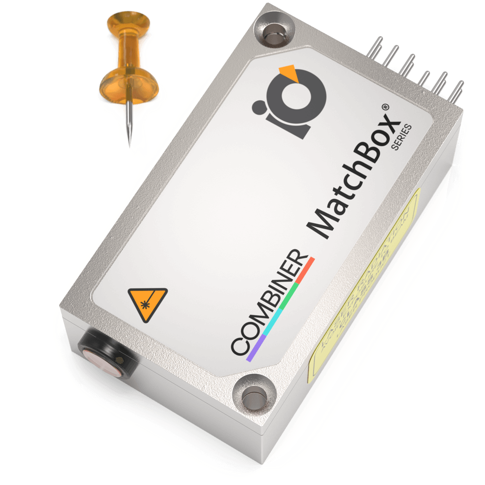
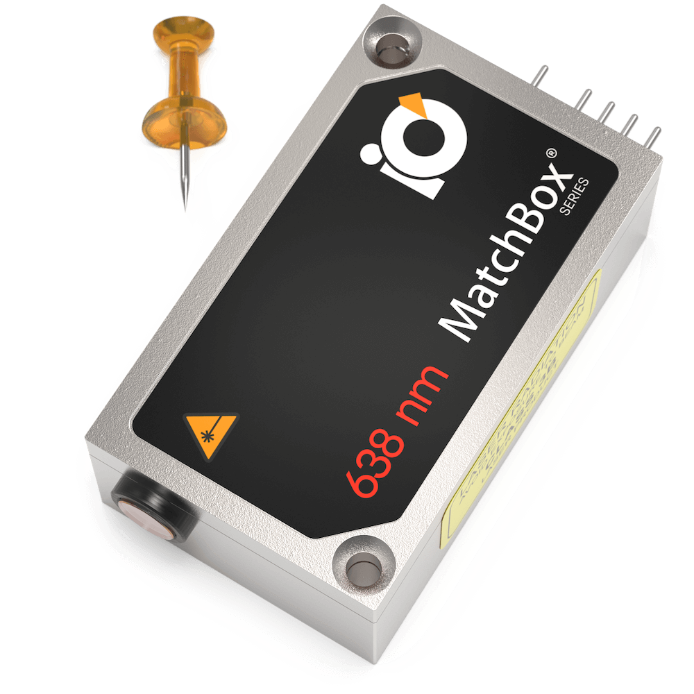
Here at Integrated Optics, we produce lasers that are perfect light sources for confocal microscopy. We have a wide range of selectable wavelengths, including the most popular choices for fluorescence and confocal microscopy (405 nm, 488 nm, 532 nm, 638 nm) with free - space or fiber-coupled outputs. While our wavelength combiners are perfect for more high-end confocal microscopy systems intended for multi-channel fluorescence detection and live cell imaging.
Relevant Products
Multi-Wavelength Laser
488 nm - 40
561 nm - 50
638 nm - 130
Power stability, % (RMS, 8 hrs): 0.2
Intensity noise, % (RMS, 20 Hz to 20 MHz): 0.5

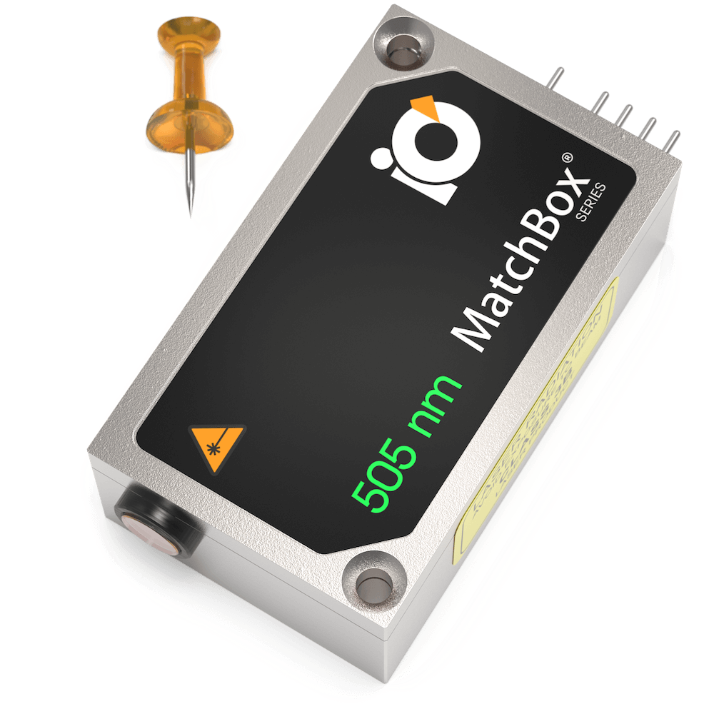
505 nm Laser
Spectral line width FWHM, nm: 0.7
Output power, mW: 60
Power stability, % (RMS, 8 hrs): 0.03
Intensity noise, % (RMS, 20 Hz to 20 MHz): 0.5
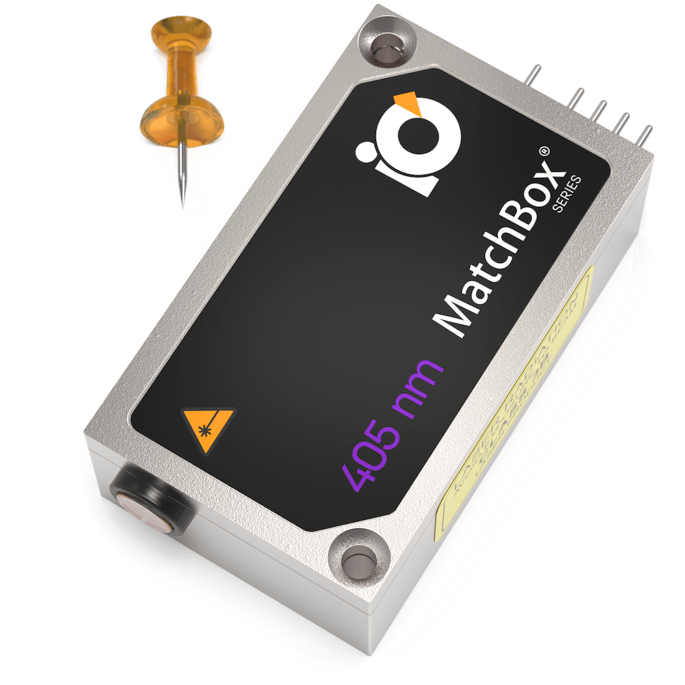
405 nm SLM Laser
Spectral line width FWHM, MHz: 20
Output power, mW: 50
Power stability, % (RMS, 8 hrs): 0.05
Intensity noise, % (RMS, 20 Hz to 20 MHz): 0.2
Multi-Wavelength Laser
488 nm - 40
520 nm - 80
638 nm - 130
Spectral line width FWHM, nm: 1
Power stability, % (RMS, 8 hrs): 0.2
Intensity noise, % (RMS, 20 Hz to 20 MHz): 0.5
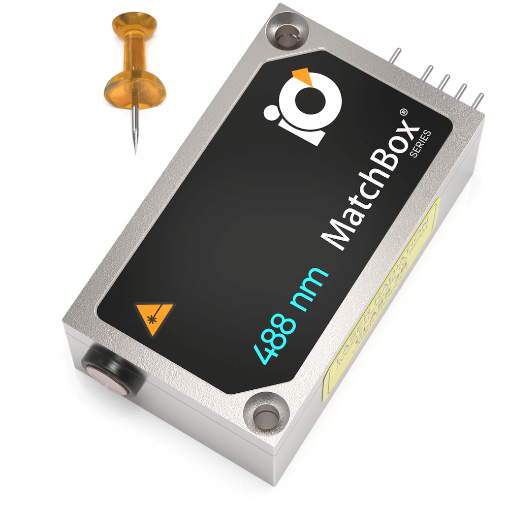
488 nm SLM Laser
Spectral line width FWHM, pm: 0.1
Output power, mW: 30
Power stability, % (RMS, 8 hrs): 0.05
Intensity noise, % (RMS, 20 Hz to 20 MHz): 0.1
633 nm SLM Laser
Spectral line width FWHM, MHz: 2
Output power, mW: 70
Power stability, % (RMS, 8 hrs): 0.03
Intensity noise, % (RMS, 20 Hz to 20 MHz): 0.2

638 nm SLM Laser
Spectral line width FWHM, MHz: 2
Output power, mW: 100
Power stability, % (RMS, 8 hrs): 0.03
Intensity noise, % (RMS, 20 Hz to 20 MHz): 0.25
Publications
ACS: The Journal of Physical Chemistry C:
Nano Letters:
Nature: Molecular Psychiatry:
Light: Advanced Manufacturing :
Small:
Biomedical Optics Express:
Sensors: